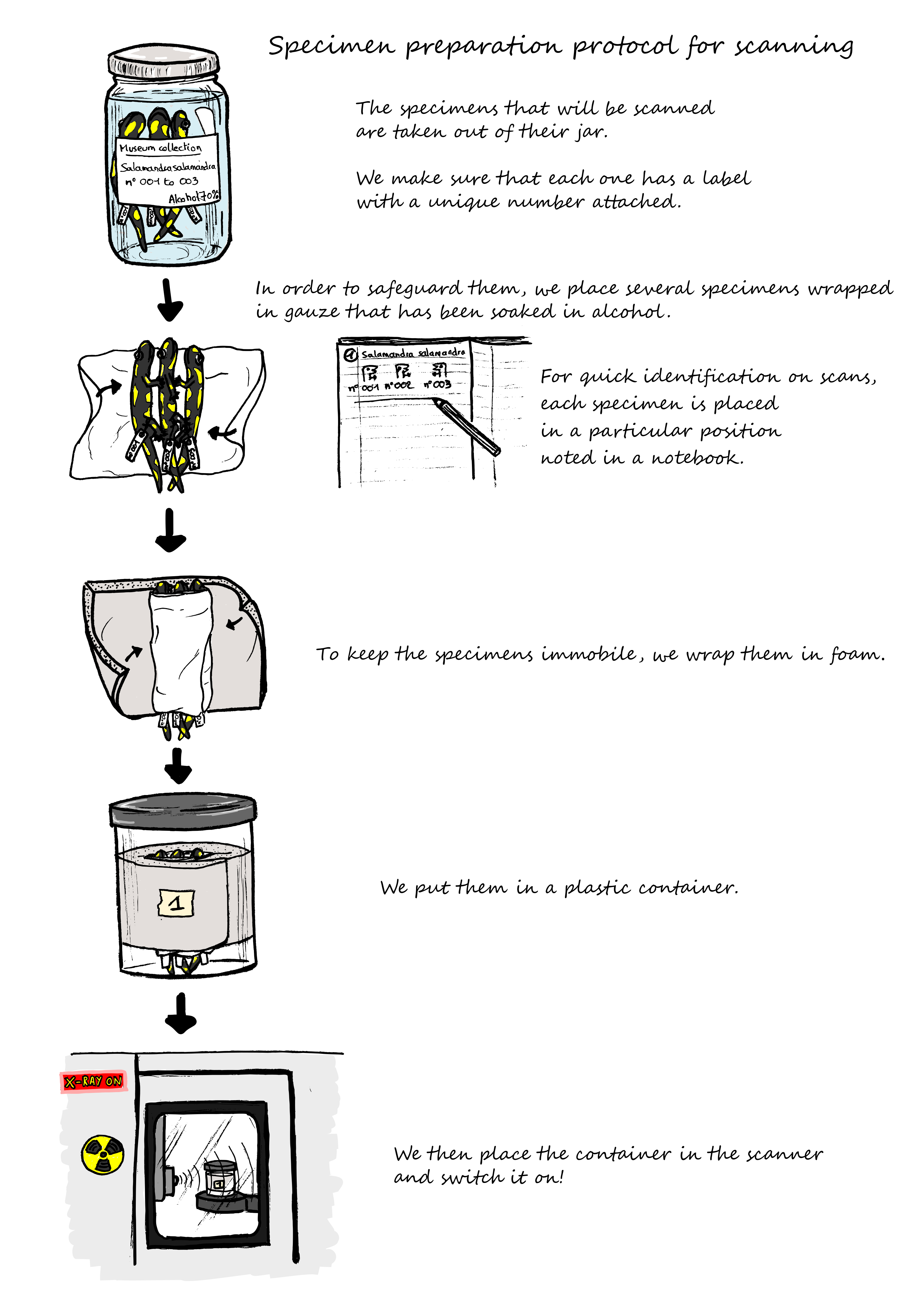
In order to observe the precise morphology of the head bones, we use a micro-computed tomography (micro-CT scanner). This is a 3D imaging technique using x-rays to see inside an object, slice by slice.
For our project, we need to study a large number of salamanders and newts, preserved in alcohol jars, in museum collections. They are are precious and must be returned to the collection in good condition.
We therefore set up a preparation protocol enabling simultaneous scanning of several specimens, while ensuring that they remained in good condition and immobile during the scanning process.