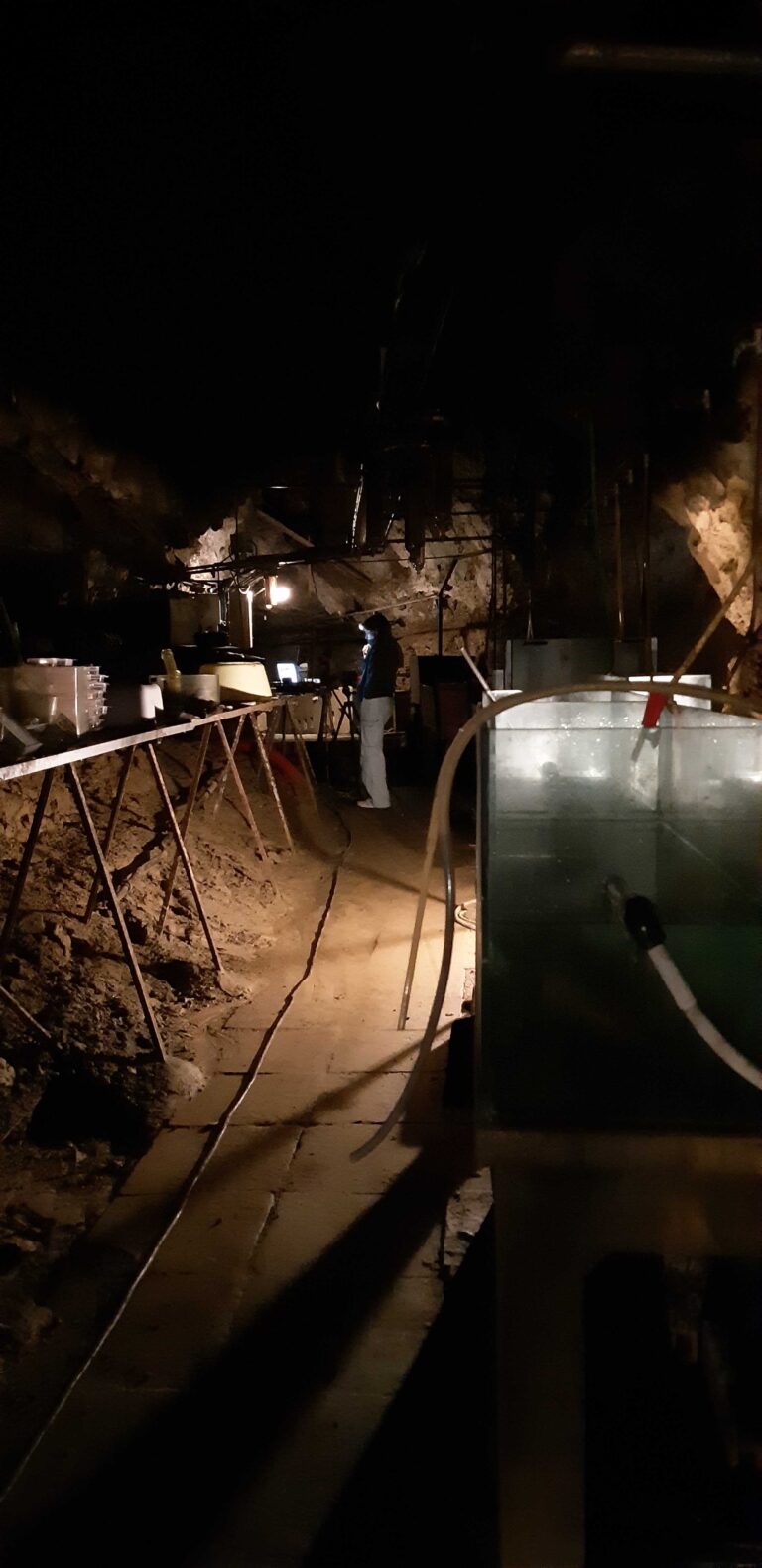
- To collect data on skull shape from salamander specimens we use CT scanning, such as with this micro CT scanner. This allows us to collect data from a specimen non-destructively so that the specimen remains preserved for future research.
- Since it can take over an hour to perform a single scan, we bundle multiple salamander specimens together in a plastic tube that will be inserted into the scanner.
- A CT scanner uses x-rays, which can pass easily through low-density materials such as cloth, foam, and plastic. So we use foam pads and other materials to pack the specimens tight to prevent the specimens from moving during the scan.
- Here's what the bundle of salamander specimens look like when viewed with x-rays. We can use an x-ray preview to make sure everything is aligned properly before starting the scan.
- Vivien viewing the results of a newly finished micro CT scan.
- Using the CT scanning software we perform a quick segmentation of the scan, which isolates the higher density bone from the lower density soft tissues and packing materials. This allows us to quickly check that the scan turned out OK.
- Micro CT scanning enables us to obtain high-resolution 3D anatomical data from the full skeleton of a salamander specimen, without damaging the specimen in any way.
CT scanning natural history collections
To collect data on the diversity of shapes and sizes in salamanders, we use micro CT scanning. In this photo gallery we share photos of a recent visit to the CT scanning facility and explain how we create high-resolution meshes from natural history specimens.