

In order to observe the precise morphology of the head bones, we use a micro-computed tomography (micro-CT scanner). This is a 3D imaging technique using x-rays to see inside an object, slice by slice.
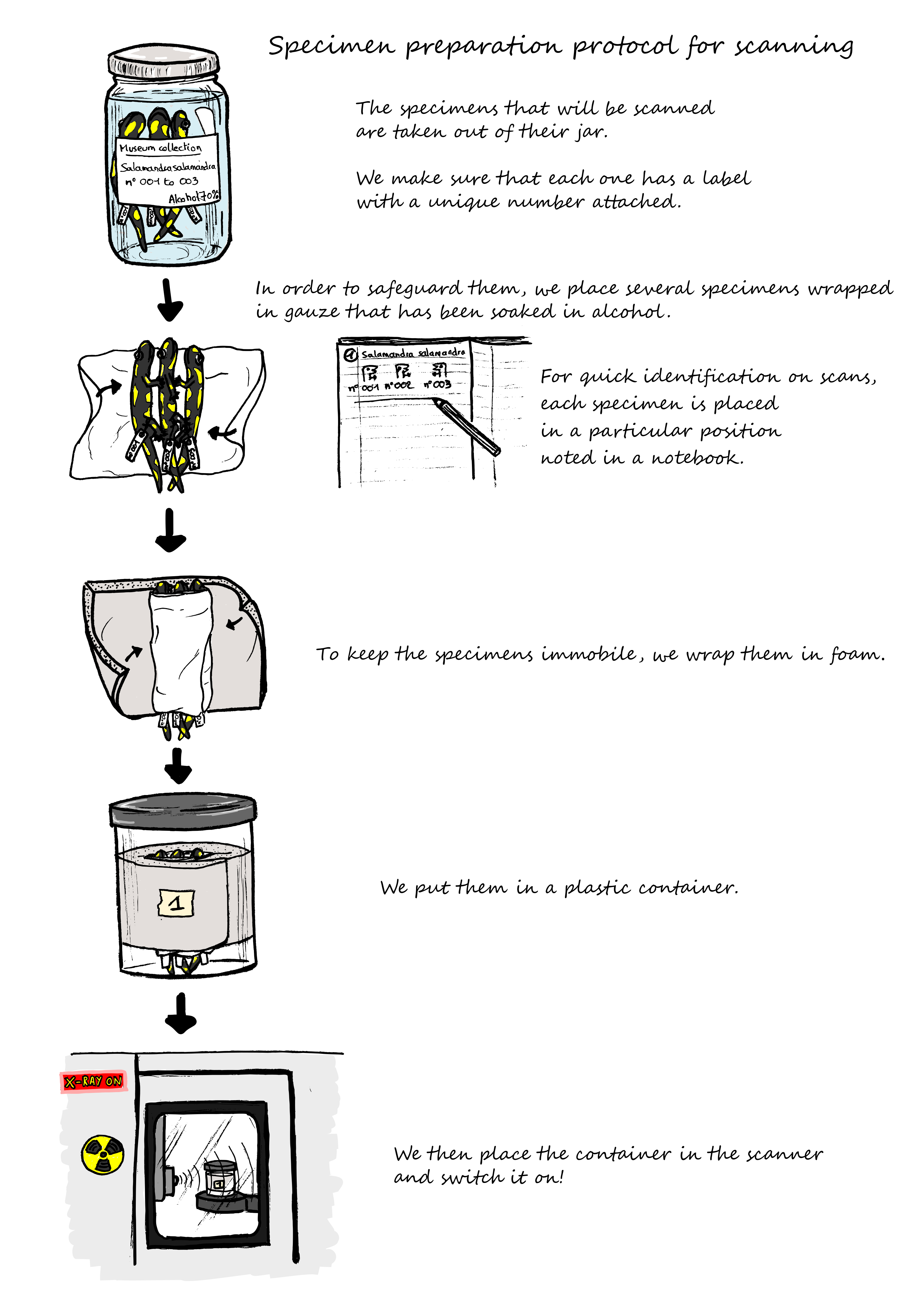
For our project, we need to study a large number of salamanders and newts, preserved in alcohol jars, in museum collections. They are are precious and must be returned to the collection in good condition.
We therefore set up a preparation protocol enabling simultaneous scanning of several specimens, while ensuring that they remained in good condition and immobile during the scanning process.
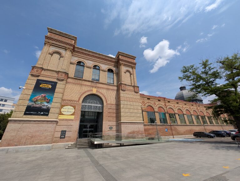
Here are a few photos of two of our CT scan missions in Tübingen and Madrid:
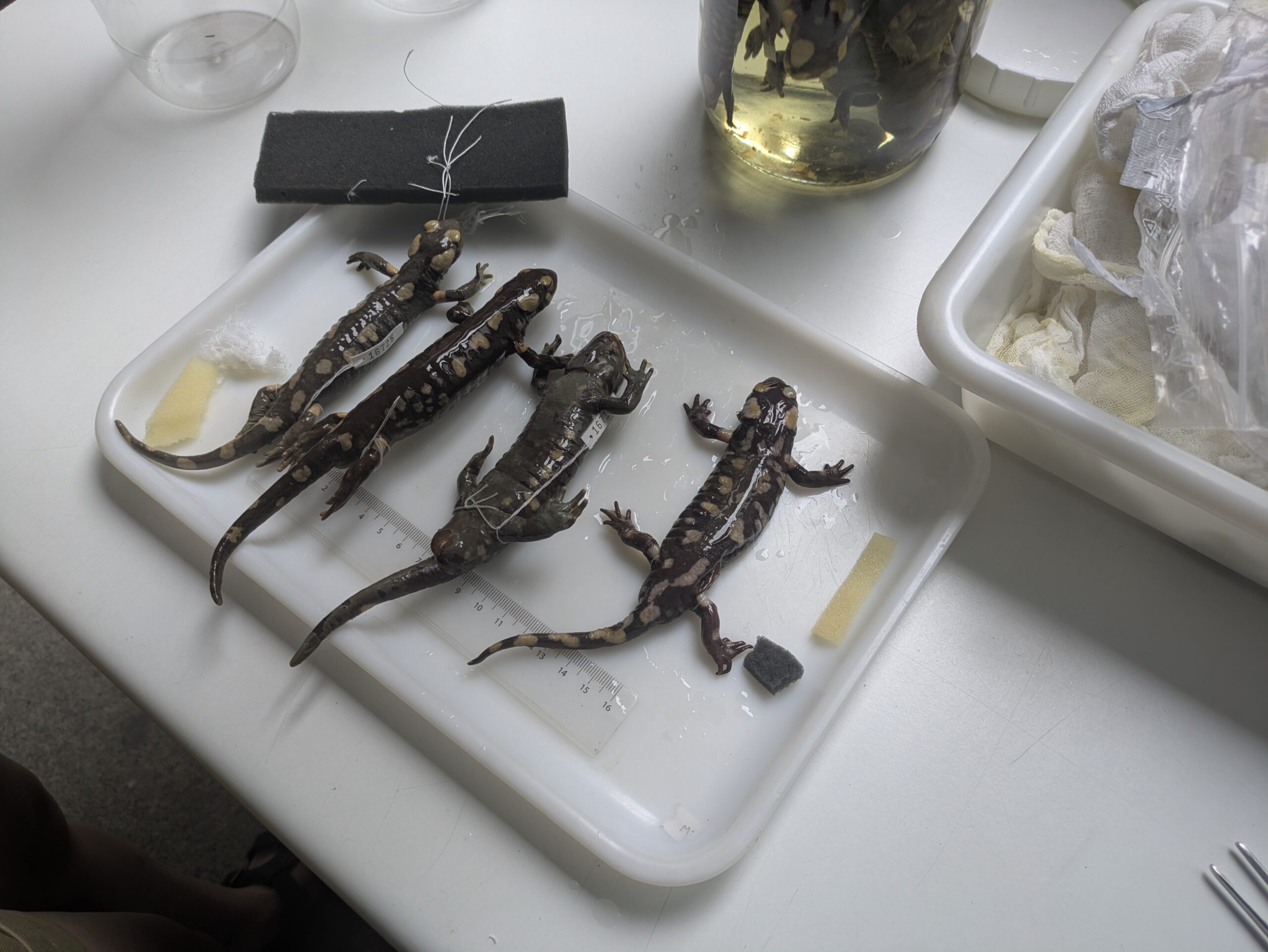

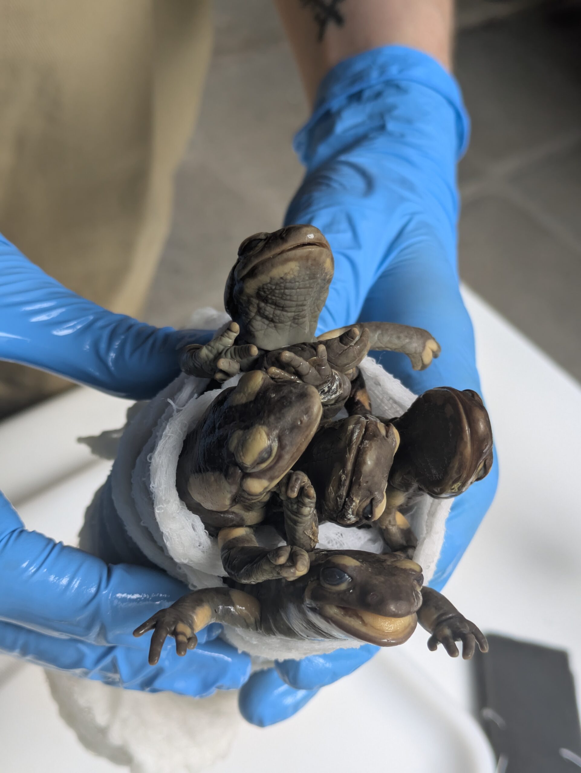

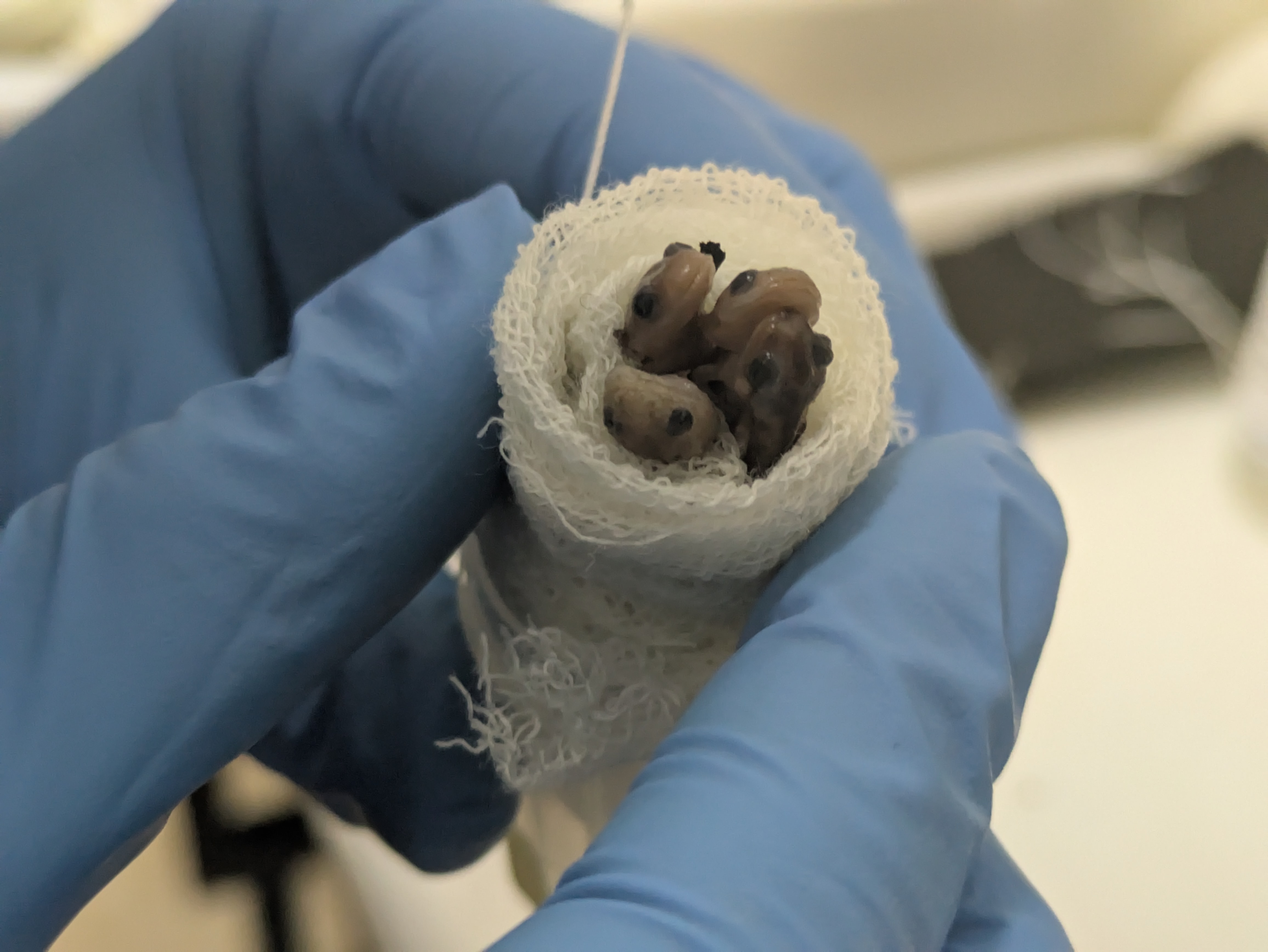

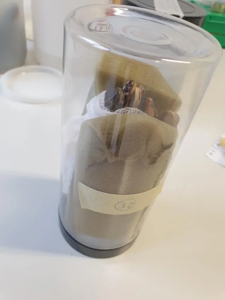

Since it can take over an hour to perform a single scan, we bundle multiple salamander specimens together in a plastic tube that will be inserted into the scanner. 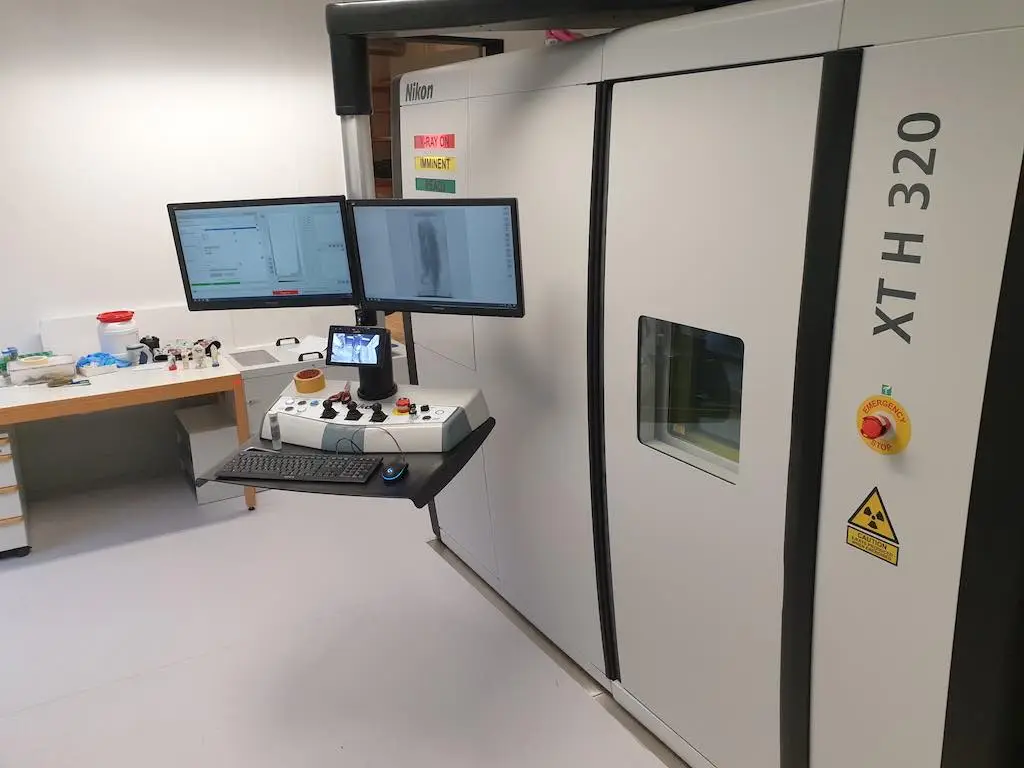

To collect data on skull shape from salamander specimens we use CT scanning, such as with this micro CT scanner. This allows us to collect data from a specimen non-destructively so that the specimen remains preserved for future research. 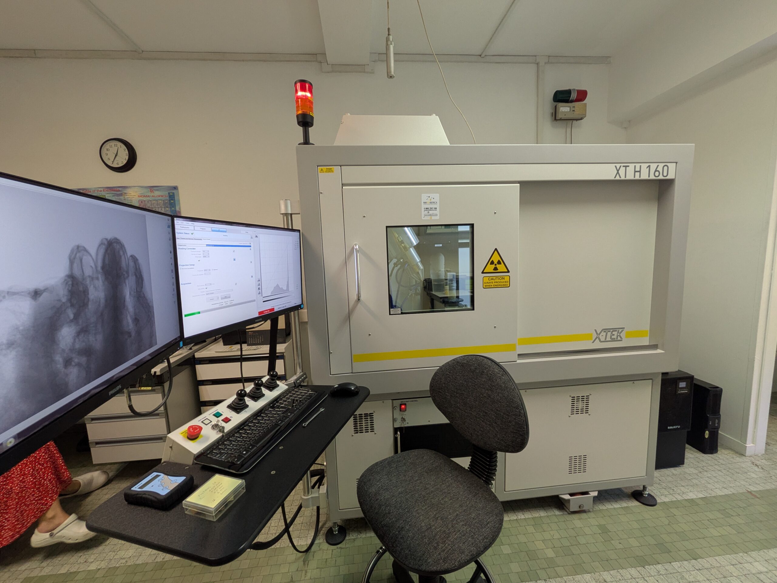
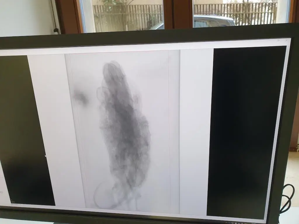
Here’s what the bundle of salamander specimens look like when viewed with x-rays. We can use an x-ray preview to make sure everything is aligned properly before starting the scan. 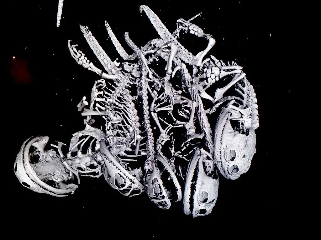
Micro CT scanning enables us to obtain high-resolution 3D anatomical data from the full skeleton of a salamander specimen, without damaging the specimen in any way.